



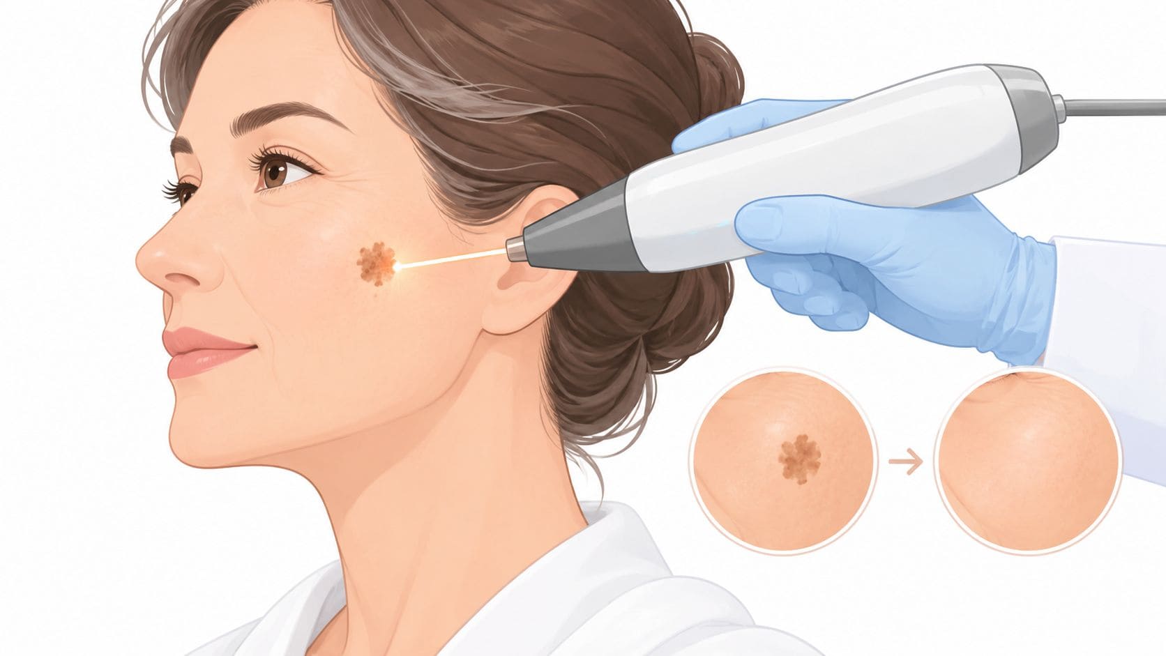
A 50-something woman in Cape Town has spent years trying fading creams on the brown marks across her cheeks and forearms. They soften briefly, then the spots seem to return after another season outdoors. In the consultation room, she asks the question many South African patients ask: will an age spot removal laser finally clear the sun spots, and will it be safe for her skin tone?
The honest answer depends on three things: whether the mark is a solar lentigo, how deep the pigment sits, and how the device and settings match the patient's phototype. South African clinics already treat pigmentation, sun damage and sun spots as mainstream dermatology and aesthetic services, with one Cape Town clinic listing age-spots removal at R350 to R950 for a 30-minute session and other practices listing pigmentation treatments from R590 to R2,310 (South African specialist clinic listings). That accessibility has made pigment treatment a repeatable outpatient service, but it hasn't removed the need for careful diagnosis and conservative technique.
Table of Contents
- How Lasers Remove Age Spots
- Comparing the Main Laser and Light Technologies
- Assessing the Right Patient for Treatment
- Treatment Protocols, Sessions and Downtime
- Risks, Recurrence and When Not to Treat
- Pre- and Post-Treatment Care That Protects Results
- Pricing, ROI and Marketing for Aesthetic Clinics
How Lasers Remove Age Spots
A patient from Cape Town presents with flat, sharply defined marks on sun-exposed skin. The pattern may match solar lentigines, the clinical term for age spots linked to cumulative ultraviolet exposure. Appearance alone cannot confirm that diagnosis. A changing, raised, irregular or bleeding lesion requires medical assessment before any cosmetic device is considered.
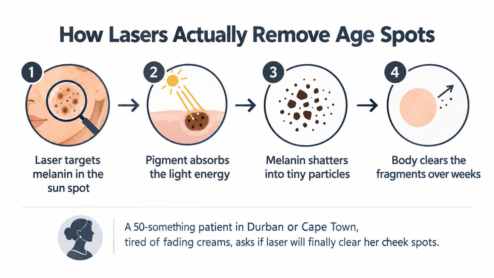
For a confirmed benign lentigo, treatment relies on selective photothermolysis. The device delivers light at a wavelength absorbed more strongly by melanin than by nearby tissue. Melanin takes in the energy, converts it into heat and fragments, while the clinician aims to limit heat reaching surrounding skin.
The pigment does not disappear during the appointment. Macrophages, immune cells that remove cellular debris, help clear the disrupted melanin over the following weeks. The mark may first look darker, followed by fine crusting or flaking, before it fades. Patients who expect an instant eraser may mistake this normal healing stage for treatment failure, so the clinic should explain the sequence before treatment.
Why pulse duration changes the safety margin
Q-switched devices deliver energy in very short pulses. Picosecond devices use shorter pulses still. Both approaches can fragment pigment rapidly, leaving less time for heat to spread into adjacent tissue. That distinction matters for darker South African phototypes, where excess epidermal heating can trigger post-inflammatory hyperpigmentation.
The European Society of Laser in Dermatology identifies ultrashort Q-switched or picosecond systems as a preferred approach for actinic lentigines. Its guidance indicates that these lesions usually clear in 1–2 sessions, with better outcomes when fluence remains conservative in darker phototypes (clinical guidance on laser treatment for solar lentigines). The figure is not a guarantee. Pigment depth, recent UV exposure, lesion type and individual healing can change the response.
Broadband IPL follows a different principle. It delivers a filtered flash containing a range of wavelengths rather than one laser wavelength. This can cover a wider field of photo-damage, but it also exposes surrounding chromophores to broader energy. Recently tanned skin and darker phototypes therefore require particular care in patient selection and settings.
Patients often use “laser” to describe every light treatment. Clinic teams should distinguish a focused pigment laser, broadband IPL and a resurfacing platform because they address different targets. This overview of what skin resurfacing does can help explain why pigment clearance and texture improvement are separate goals.
A brown patch is not automatically suitable for pigment treatment. Facial redness or flushing may point to another diagnostic pathway, including treating rosacea with a dermatologist, before the clinic considers a light-based procedure. Clear explanations protect patients and help clinics build repeatable, trust-based treatment plans around South Africa's strong UV exposure.
Comparing the Main Laser and Light Technologies
A clinic owner comparing devices should begin with the patient's pigment, not the newest handpiece. Wavelength helps determine where energy is absorbed. Shorter wavelengths act closer to the surface, while longer wavelengths can reach deeper pigment. Superficial epidermal pigment generally responds to energy in the 500–600 nm range. Deeper or dermal pigment may require a longer wavelength such as 1064 nm (wavelength and pigment-depth guidance).
The main technologies serve different jobs:
- IPL uses filtered broadband light across a range of wavelengths. It can treat broader areas of superficial, sun-related colour change when the patient, lesion and skin condition are suitable.
- Q-switched Nd:YAG commonly uses 1064 nm for deeper penetration and frequency-doubled 532 nm for more superficial pigment.
- Picosecond systems may offer 532, 755 or 1064 nm options. Their very short pulses concentrate energy in the pigment while limiting heat spread.
- Fractional systems create microscopic treatment zones for texture and wider photo-damage. Ablative wavelengths include CO2 at 10 600 nm and Er:YAG at 2945 nm. Non-ablative systems may use 1550 or 1927 nm.
The table is a clinical orientation, not a promise of clearance or a substitute for a test spot. Session requirements, safety limits and fees vary with the lesion, device, operator and protocol. A South African clinic should complete its pricing column from audited local fees rather than treating a general table as a quotation. A local pricing reference can help the team check how fees are presented in practice (local pricing reference).
| Technology | Wavelength / Mechanism | Best lesion type | Sessions needed | Fitzpatrick safe range | Downtime | Cost per session (ZAR) |
|---|---|---|---|---|---|---|
| IPL | Filtered broadband light, commonly covering a broad range such as 515–1200 nm | Diffuse superficial photo-damage and suitable superficial sun spots | Individual plan required | More predictable in lighter, untanned phototypes. Caution rises in IV–VI | Redness and darkening may occur | Use local clinic pricing |
| Q-switched Nd:YAG | 532 nm for superficial pigment, 1064 nm for deeper penetration, short pulses | Discrete lentigines and selected deeper marks | Often planned as a course when needed | 1064 nm is generally the more conservative option for darker phototypes | Short inflammatory phase, possible crusting | Use local clinic pricing |
| Picosecond | 532, 755 or 1064 nm, very short pulses that limit heat spread | Resistant discrete pigment and selected darker-phototype cases | Individual plan required | Can be useful across a broad range, but still needs conservative settings and testing | Usually limited to the treated sites | Use local clinic pricing |
| Fractional laser | Ablative or non-ablative microscopic treatment columns | Texture-led photo-damage with associated uneven colour | Individual plan required | Ablative approaches require particular caution in IV–VI | Non-ablative recovery is usually lighter. Ablative recovery is longer | Use local clinic pricing |
The practical choice depends on the pattern being treated. Superficial sun spots on lighter, untanned skin may suit IPL or 532 nm. Deeper pigment points towards 1064 nm. Resistant lesions or darker phototypes may favour picosecond treatment with conservative settings and test areas. Texture-led photo-damage with pigment may be better suited to fractional non-ablative treatment than to a spot-only laser.
South African UV exposure makes this distinction useful in patient conversations. A device that clears a visible mark does not remove the reason new pigment may appear. Clinics should therefore explain why maintenance, assessment and appropriate follow-up may form a repeatable care plan, while avoiding promises about permanent clearance.
Teams considering Nd:YAG capability should understand the difference between wavelength options and lesion depth. This guide to YAG laser treatments offers a practical starting point for explaining the technology without presenting every brown mark as the same indication.
Assessing the Right Patient for Treatment
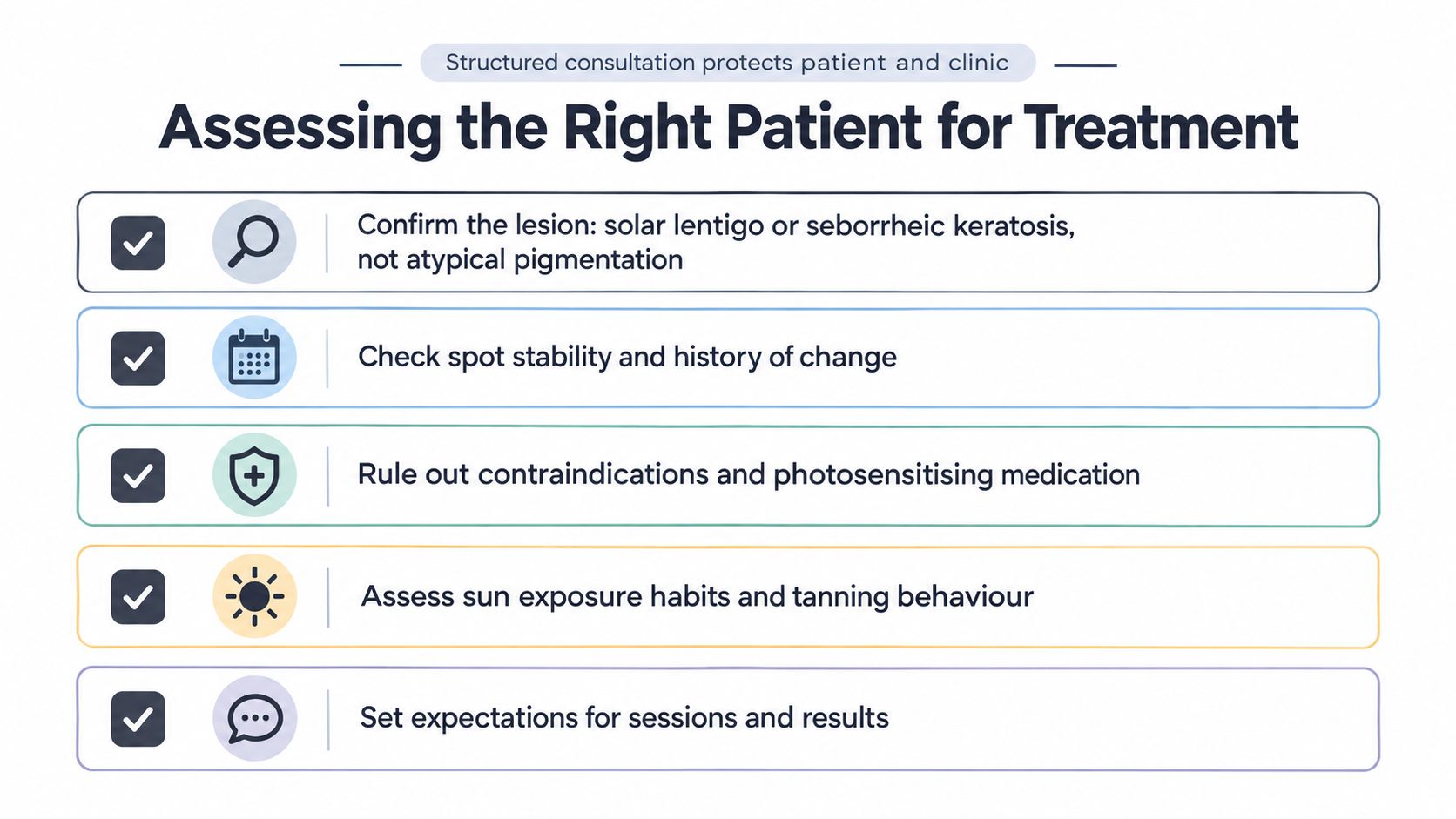
A patient arrives asking to remove one brown mark before a holiday. The consultation should begin with the lesion, not the device. First establish what the mark may be, then decide whether energy-based treatment is appropriate.
Examine it closely and record its history. A flat, stable, evenly coloured mark may fit a solar lentigo. A raised or scaly lesion may be a seborrheic keratosis, while an atypical pigmented lesion may need dermatology referral. Document borders, colour variation, size and any change over time. Use the ABCDE framework as a screening prompt, not as a diagnosis.
The consultation questions that change the plan
Ask about:
- Pigment characteristics: Is the pigment epidermal or dermal, sharply bordered or diffuse, isolated or widespread?
- Skin response: What is the patient's Fitzpatrick phototype? Have peels, waxing or devices previously caused darkening?
- Recent UV exposure: Has the patient been outdoors, tanning or sunburnt recently?
- Medication: Are isotretinoin, photosensitising medicines or immunosuppressive treatments involved?
- Hormonal and medical context: Could pregnancy, melasma, hormonal pigmentation or a history of keloids affect the plan?
- Current skincare: Are retinoids, hydroquinone, exfoliating acids or other sensitising actives being used?
South African clinics need particular care because many patients have phototypes IV–VI. Wavelength and fluence must suit the patient's melanin, or post-inflammatory hyperpigmentation can replace the original concern. The linked clinical guidance also warns that outcomes are less predictable in skin of colour and that permanent hypo- or depigmentation can occur after treatment.
Practical candidacy rules
Phototypes IV–V may be candidates for conservative 1064 nm Q-switched or picosecond treatment, while aggressive IPL is a poor default. Phototype VI requires especially cautious parameters, test spots and close follow-up. Referral is sometimes the safest decision.
A raised, changing, painful or bleeding lesion should not be lasered in an aesthetic setting. Pigmented lesions should be diagnosed appropriately before treatment. Once suitability is established, Q-switched Nd:YAG may be considered for tan, blue-grey or brown marks at varying depths (diagnosis before pigmented-lesion treatment).
For a clinic, the assessment form supports clinical reasoning as well as administration. It records why the lesion appeared suitable, captures risk factors, supports consent and gives the team a defensible basis for choosing a test spot, referral or alternative treatment. This guide to treating pigmentation may support staff education, but it does not replace examination by an appropriately qualified clinician. In a high-UV setting, careful selection also supports honest discussions about maintenance and recurring care, rather than promises of permanent clearance.
Treatment Protocols, Sessions and Downtime
A patient arrives expecting one quick treatment, while the clinic sees a lesion whose depth, skin tone and response still need to be assessed. The treatment timeline should make that uncertainty clear. A session plan is a working framework, not a promise of complete clearance.
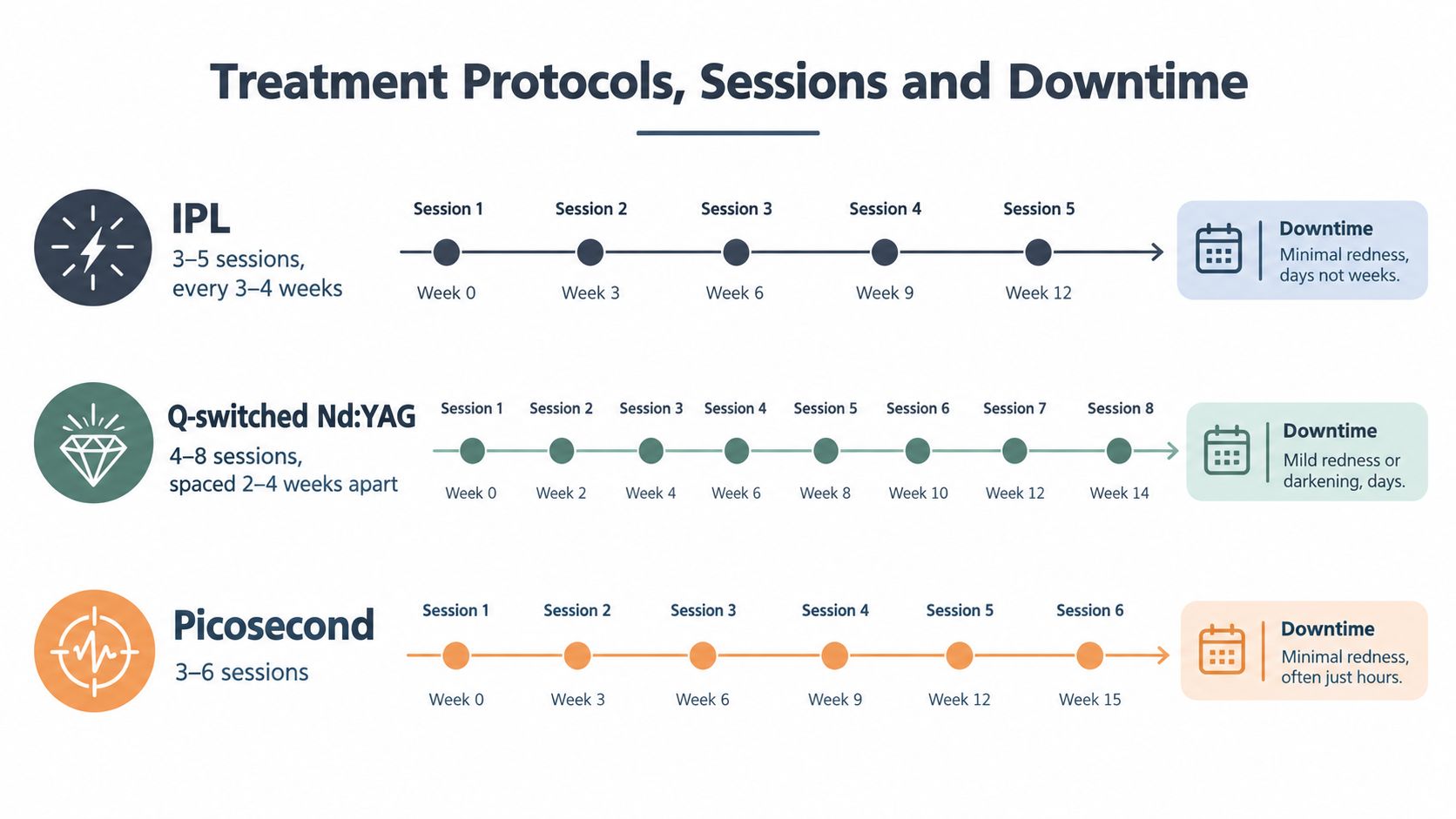
Common planning ranges include:
- IPL: often 3–5 sessions at 3–4 week intervals.
- Q-switched Nd:YAG: often 4–8 sessions spaced 2–4 weeks apart.
- Picosecond: often 3–6 sessions at monthly intervals.
- Fractional non-ablative: often 4–6 sessions.
- Fractional ablative: may be considered in 1–3 sessions, followed by a longer recovery period.
These ranges must be adapted to the device, indication, treated area and patient response. A clinic should check its device manual, medical oversight and treatment records before presenting a protocol. For South African patients, UV exposure and higher epidermal melanin can make conservative progression more appropriate, particularly when the clinic is building a maintenance plan rather than selling a single clearance session.
What patients usually see
Immediately after pigment-targeting treatment, a spot may whiten, redden or swell slightly. During the following days, it may darken and form microcrusting. Flaking often occurs between days 5 and 10, and visible fading may be easier to judge during weeks 2–4.
Explain the process using a simple comparison: the treated pigment behaves like fine debris being cleared gradually, not like ink disappearing at the moment the laser fires. Patients should leave crusts alone. Excessive pain, blistering, spreading redness or an unexpected colour change requires prompt review.
The face may appear to respond sooner than the backs of the hands or the decolletage. Hands and chest receive continuing UV exposure and may show more established photo-damage, so they often need greater patience and stronger maintenance habits.
Measure the result, not memory
Take standardised photographs at every visit, using the same lighting, distance, camera position and background. Patients may focus on the darkest spot and miss broader improvement. Clinics can also overestimate progress when images differ.
Record fluence, spot size and pulse duration in the treatment note. The safest setting is the one that produces an appropriate response while respecting epidermal melanin and the patient's healing pattern. Consistent records help the team refine later sessions and explain why maintenance may support longer-lasting results.
Risks, Recurrence and When Not to Treat
A patient returns with a lighter brown spot, pleased with the immediate change. The skin is also blistered, tender and becoming darker around the treated area. Clearance alone is not a successful outcome. The clinic must assess healing, pigment change and the possibility that new spots will appear as South African UV exposure continues.
The main risks are post-inflammatory hyperpigmentation, especially in phototypes IV–VI, hypopigmentation, blistering and scarring. IPL can darken pigment paradoxically if energy is too high or the patient's phototype is unsuitable. Ablative settings create more thermal injury, so an incorrect choice can produce a longer and more visible recovery.
Recurrence counselling needs local context. A South African review examined gingival pigmentation rather than facial solar lentigines, so its findings cannot be transferred directly to age spots. It reported repigmentation of 22.8% with laser therapy versus 21.4% with scalpel treatment, while one laser modality showed up to 30% recurrence within six months (South African review of laser treatment and repigmentation). The figures do not predict a facial result. They do show why removing visible pigment does not prevent future pigment biology or UV-related change.
| Laser type | Phototypes I–III | Phototypes IV–VI |
|---|---|---|
| IPL | May suit selected superficial, high-contrast pigment when the skin is not recently tanned | Epidermal melanin raises the risk of post-inflammatory hyperpigmentation. Select carefully or avoid |
| 532 nm Q-switched | Allows precise targeting of suitable superficial pigment | Use conservative fluence, test spots and close monitoring |
| 1064 nm Q-switched Nd:YAG | May suit deeper pigment or an indication that supports this wavelength | Often the more cautious wavelength choice, though it still carries risk |
| Picosecond | May suit concentrated or resistant pigment | A selective option may be possible, with test treatment and conservative parameters |
| Fractional ablative | May address texture alongside deeper photodamage | Prolonged inflammation and pigment change are more likely. Specialist protocols are required |
| Fractional non-ablative | May suit texture and diffuse photodamage | Careful parameter selection and aftercare remain necessary |
When the correct decision is no treatment
Postpone treatment for an active tan, open lesion, suspected melanoma or mark with concerning ABCDE features. Pregnancy, recent isotretinoin use, significant immunosuppression and some photosensitising medicines may require postponement, medical review or referral. A history of keloids, melasma overlap, hormonal pigmentation or recent filler in the treatment zone also warrants caution.
Use dermoscopy or refer to dermatology when the diagnosis is uncertain. If a lesion appears suitable but the response is difficult to predict, especially in a darker phototype, begin with a single low-fluence test spot. IPL is not a default choice for every brown mark. A longer-wavelength Nd:YAG approach may reduce epidermal melanin exposure, but it is not risk-free.
Maintenance discussions can support ethical recurring care. Explain that treated spots and newly formed spots are separate issues, rather than promising permanent protection from UV damage. Repeated sessions should follow a documented clinical reason, not an automatic sales cycle.
Pre- and Post-Treatment Care That Protects Results
A patient arrives for age spot removal with a fresh tan, irritated skin and several active products still in use. The device may be suitable, yet the starting conditions increase uncertainty. Good aftercare begins before the first pulse by keeping the skin calm, untanned and free from avoidable irritation. This reduces competing inflammation and limits UV-driven pigment during recovery.
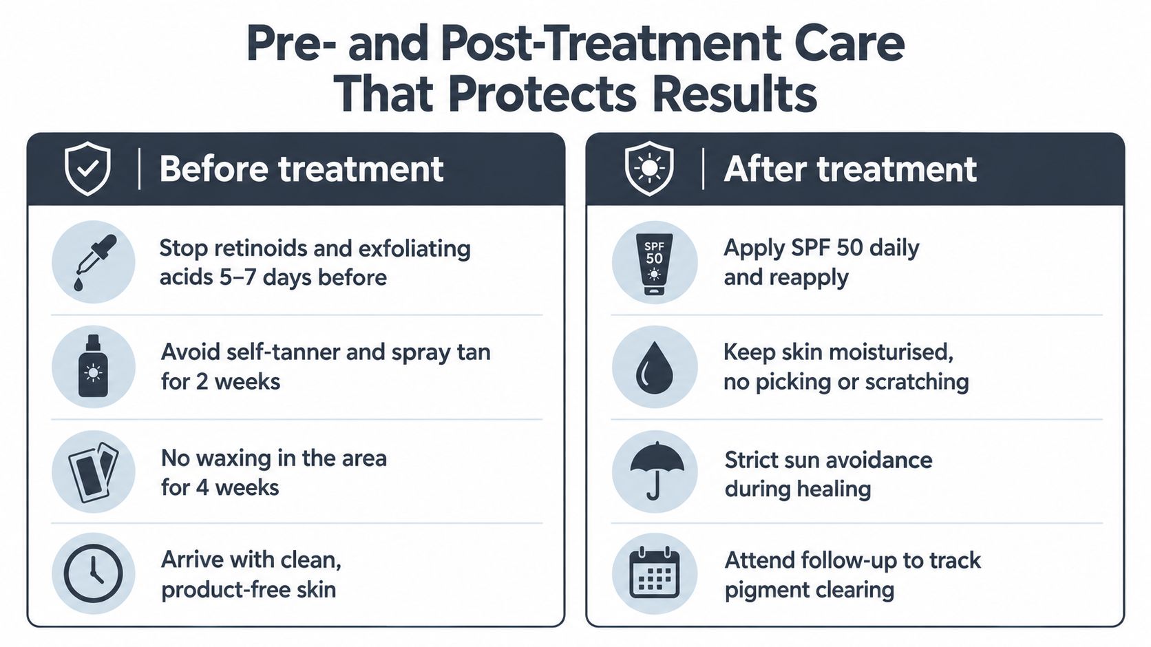
Before the appointment
A clinic handout should give patients clear actions:
- Active skincare: Pause retinoids and exfoliating acids 5–7 days before treatment, unless the treating clinician provides different instructions.
- Artificial colour: Avoid self-tanner and spray tan for 2 weeks.
- Waxing: Avoid waxing the treatment area for 4 weeks.
- Sun protection: Apply SPF 50+ with strong UVA protection before and after treatment. South African sun-protection guidance for procedures recommends strict protection for 2–4 weeks before treatment and continuation for 2 weeks after inflammation resolves.
- Appointment preparation: Arrive with clean, product-free skin and disclose every medicine and active product.
Retinoids and acids can increase irritation, while waxing can disturb the surface barrier. Tanning adds epidermal melanin that competes with the target for the device's energy. In practical terms, the clinician needs a predictable canvas, particularly when treating darker South African skin tones in a high-UV environment. A list of pre- and post-treatment care instructions for skin procedures such as laser treatments.
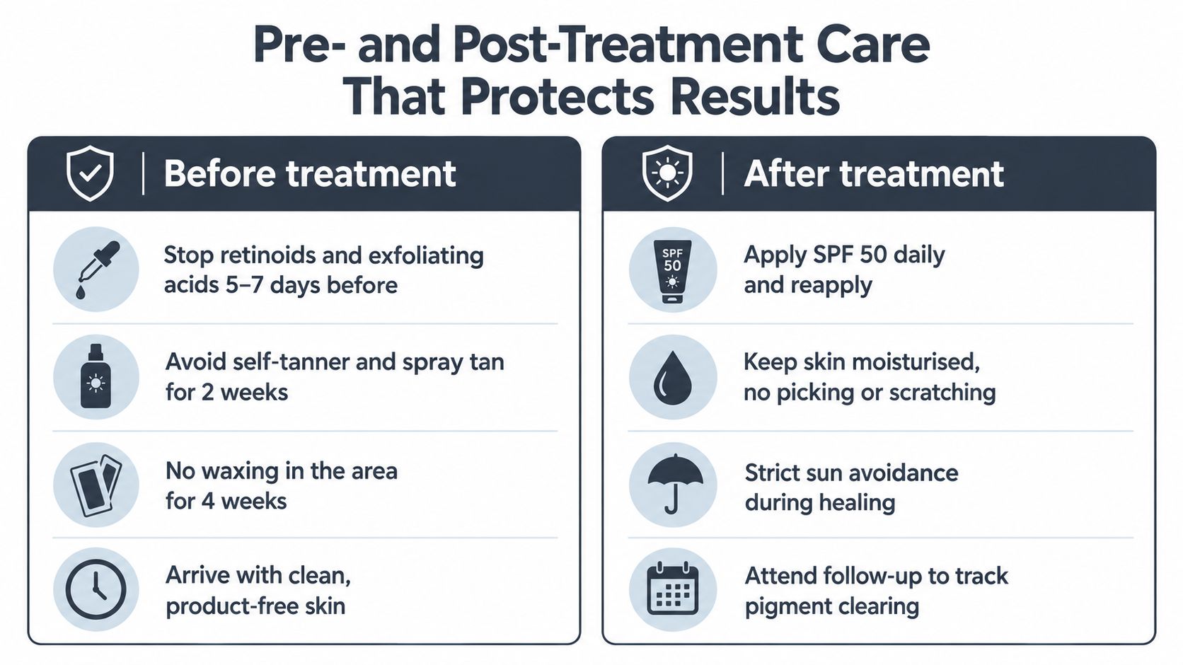
After treatment
For the first 24 hours, cool compresses can ease warmth and swelling. A plain emollient or petrolatum-based barrier may suit ablative treatment, according to the clinician's protocol.
For 48–72 hours, follow the provider's advice about avoiding picking, scratching, heat, saunas and strenuous exercise. Do not restart active ingredients until the skin has re-epithelialised and feels comfortable. During healing, use strict SPF 50 protection, with physical blockers where appropriate, and follow the clinic's stated period of sun avoidance.
A printable timeline helps reception staff reinforce the same advice:
- Day 1: Cool the area, keep products simple and monitor the response.
- Week 1: Darkening, microcrusting or flaking may occur. Do not pick.
- Week 4: Review fading, compare standardised photographs and confirm whether the skin is ready for another session.
- Week 12: Assess the broader result, discuss prevention and decide whether maintenance has a clinical reason.
Melanin remains responsive to environmental stimulation after treatment. UV exposure can encourage new pigment before the body has finished clearing disrupted material, reducing visible improvement and increasing uneven colour. Explaining this distinction helps patients understand why sun protection protects both the immediate result and the clinic's longer-term treatment plan.
Pricing, ROI and Marketing for Aesthetic Clinics
A patient arrives after noticing a dark patch before a wedding. The mark may be suitable for laser or light treatment, but the appointment also carries diagnostic, photographic, counselling and follow-up work. A sustainable service prices that full pathway, not only the device's pulses.
South African pricing gives clinics a useful local reference. Local listings span R350–R2,310 depending on treatment area and modality (South African clinic pricing reference). That spread supports publishing a clear per-area price list rather than benchmarking against one provider or promising a universal price for IPL, Q-switched, picosecond or fractional treatment. The final fee should reflect the patient's indication, skin type, treatment area, operator time and review plan.
| Technology | Per-Session Price Band (ZAR) | Typical 3-Session Package | Gross Margin Range | Breakeven Context |
|---|---|---|---|---|
| IPL | Set from local treatment-area pricing and operating costs | Offer only where three sessions are clinically appropriate | Calculate from actual labour, consumables and room costs | Review utilisation, filter or lamp costs and treatment time |
| Q-switched Nd:YAG | Set by spot count, area and operator time | Useful for a defined course, with review before commitment | Calculate from shot use, maintenance and staff time | Factor device finance, service costs and treatment throughput |
| Picosecond | Set according to device investment and clinical complexity | Use staged packages with reassessment | Calculate from pulse consumption, training and room use | Require enough suitable cases to support the capital cost |
| Fractional treatment | Set according to area, downtime and operator skill | Package only when the indication supports a course | Calculate from consumables, recovery support and appointment length | Include downtime between bookings and aftercare workload |
Build the model from real clinic inputs
Breakeven starts with fixed costs, including device finance, service agreements, insurance, compliance and training. Add variable costs such as consumables, protective equipment, topical anaesthesia where used and replacement parts.
Capacity matters just as much. Record treatment duration, room utilisation, staff availability and follow-up appointments. Then measure clinical conversion, meaning the proportion of consultations suitable for treatment rather than the number of enquiries. A recurring service can include appropriate maintenance, skincare support and referrals, but it should never depend on unnecessary sessions.
A three-session bundle can make payment easier, provided the consultation leaves room to change course. A patient may need referral after assessment, or may respond well enough that another session is not justified. Packages should reward commitment without turning a provisional estimate into a guarantee.
Market the problem patients recognise
For South African patients, high-UV messaging can focus on sun-damage reversal, even-looking skin and realistic downtime. Avoid flawless or permanent claims. A useful consultation script sounds like this:
Consultation language: “Your mark appears suitable for assessment as a solar lentigo, but we need to confirm that before treatment. The device can break down visible pigment, while sun protection helps reduce new or returning colour. I'll explain the likely course, the healing phase and the risks for your skin type before you decide.”
Omega Lasers supplies multi-technology platforms for aesthetic practices, including systems designed for facial rejuvenation and discoloured skin, alongside training, technical support, marketing support and business-development guidance. A clinic evaluating an age spot removal laser should ask whether the platform matches its intended indications, whether staff can operate it safely, and whether the business can support diagnosis, follow-up and patient education.
Seasonal campaigns can address concern after summer or before wedding events, while allowing time for assessment and healing. The offer should be a defined consultation, an appropriate device pathway, transparent pricing and follow-up that protects the result. That approach supports patient trust and gives the clinic a clearer basis for repeat care when maintenance has a clinical reason.
Omega Lasers can help clinics evaluate multi-technology platforms, training, technical support and the operational support needed to deliver pigment and skin-rejuvenation services responsibly. Visit Omega Lasers to explore device options and build an age spot treatment pathway suited to your team, patient mix and South African practice.