




A compelling treatment photo can still mislead. A single polished image says little about skin type, treatment settings, session count, recovery, lighting, or whether the result lasted. A more useful record shows the same person at baseline, during recovery, and at a clinically meaningful follow-up, with the operator's notes beside the images.
South Africa already has a substantial market for visible, repeatable aesthetic outcomes. A published review of ISAPS data recorded 85,869 aesthetic procedures in South Africa, including 48,486 non-surgical e 37,383 surgical procedures, while facial rejuvenation included 3,414 non-surgical skin-tightening treatments e 1,149 chemical peels. The report also recorded 42,757 injectables e 29,643 botulinum toxin procedures, reinforcing the commercial importance of results that clients can see and assess (South African aesthetic procedure market data).
The gallery below treats before and after results as evidence rather than decoration. Each case should connect indication, complexion, parameters, sessions, downtime, side effects, follow-up timing, objective measures, consent, consistent photography, and clinician commentary. Where relevant, Omega Lasers systems such as EM Pulse, Derma Pro, SkinSkan Pro, and Tetra Cool can be considered according to their stated treatment categories, but a device name never replaces diagnosis or professional judgement. For topical pigmentation context, clinics can also point clients towards this guide to mandelic acid before and after results.
Índice
- 1. Laser Hair Removal Full-Body Treatment Series
- 2. Skin Rejuvenation and Texture Improvement
- 3. Acne and Active Breakout Treatment Outcomes
- 4. Pigmentation Correction and Melasma Sun Damage Treatment
- 5. Vascular Lesion Treatment for Rosacea Telangiectasia and Spider Veins
- 6. Anti-Ageing and Wrinkle Reduction Through Collagen Induction
- 7. Scar Revision for Atrophic and Hypertrophic Scars
- 8. Skin Tightening and Laxity Improvement in Face and Neck
- Before & After Results: 8 Aesthetic Treatment Comparisons
- How to Read Before-and-After Results Responsibly
1. Laser Hair Removal Full-Body Treatment Series
Before and after photography can document treatment performance only when it is tied to a controlled series of visits. The record should track hair density, shaft thickness, regrowth speed, and shaving frequency across each treated zone. It should also identify complexion, hair characteristics, treatment parameters, intervals between sessions, downtime, side effects, and the clinician's assessment.
Build the case record before judging the result
A multi-wavelength platform such as Omega Lasers' Impulso EM may be considered when the plan requires different wavelengths for varied hair and skin characteristics. Record the wavelength, fluence, pulse width, cooling method, treated zones, shaving instructions, and any missed or delayed appointments. Show legs, underarms, bikini line, back, chest, chin, or upper lip only when those areas were treated.
Use identical camera settings, distance, lighting, background, positioning, and hair-growth preparation at every timepoint. Baseline images should be followed by interim regrowth photographs and a final follow-up taken after the planned observation period. Written informed consent should define approved image use, while clinical files should be stored securely under the practice's applicable privacy obligations.

Objective measures make the comparison more useful. A clinic can record the proportion of visible regrowth in a marked area, the number of days before shaving is needed, and changes in hair calibre. These observations should sit beside the client's reported comfort and satisfaction, not replace them. A smoother image taken immediately after shaving does not demonstrate reduced follicular regrowth.
Clients also need clear advice about sun exposure, aftercare, variable response between body sites, and possible short-term reactions. The number of laser hair-removal sessions clients may need should be discussed as a planning consideration, not a fixed promise.
Market context can support service planning, but it cannot validate an individual outcome. A 2019 industry document discussed South Africa's energy-based aesthetic device market outlook (South African energy-based aesthetic device market outlook). That historical source should not be used to substantiate later market-year figures. For clinics, the commercial case is stronger when demand is paired with consistent documentation, realistic counselling, and records that make treatment response easier to assess.
2. Skin Rejuvenation and Texture Improvement
Before-and-after photographs can suggest smoother skin, but they cannot prove structural improvement without controlled documentation. Glow, hydration, makeup, and lighting may alter the image. Each case should therefore record the primary concern, skin type, active conditions, treatment modality, energy settings, treatment count, downtime, and recovery effects such as redness, swelling, tightness, tenderness, or peeling.
Fractional resurfacing and multi-modality facial treatments may target fine lines, acne scarring, enlarged pores, and uneven texture. Derma Pro may fit a clinician-led facial rejuvenation or resurfacing plan. Its presence in a case study should explain the selected protocol and expected trade-offs, not imply an equivalent to an invasive procedure.
Compare texture at the right time
Photographs taken soon after treatment may capture inflammation rather than improvement. Later images provide a better basis for reviewing surface smoothness, scar edges, pore visibility, and overall tone. Polarised or cross-polarised photography can help document colour and texture changes when the clinic keeps the camera distance, magnification, lighting, facial expression, and angle consistent.
A useful gallery links each image pair to objective observations. Clinicians can record a texture rating, visible scar depth, pore prominence, or defined treatment areas, while noting the assessment method used. These measures are not interchangeable with the client's satisfaction. A client may prioritise radiance or easier makeup application, whereas the clinician may assess texture, scar depth, or uneven pigmentation. Both perspectives belong in the record.
Recovery belongs in the gallery. Clinical evidence on early laser resurfacing recovery found that more than half of patients experienced immediate tightness, swelling, tenderness, discomfort, or burning, while later outcomes and quality-of-life scores improved (clinical evidence on early laser-resurfacing recovery). Showing only the final image omits a material part of the treatment experience.
Sun protection and gentle skincare shape interpretation, particularly for darker complexions where inflammation may be followed by pigmentary change. Clinician notes should explain why the settings were chosen, document symptoms and downtime, and state when normal skincare was resumed.
3. Acne and Active Breakout Treatment Outcomes
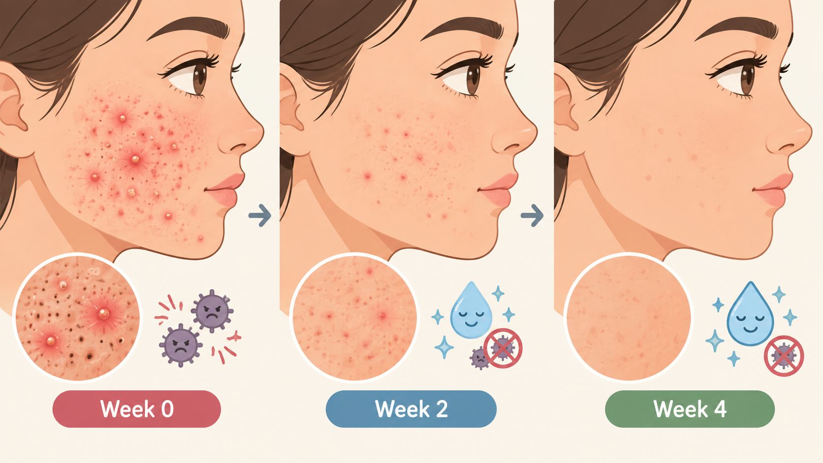
A clearer follow-up image does not prove that active acne is controlled. Reliable comparison separates inflammatory lesions, comedones, post-inflammatory marks, oiliness, and new-breakout frequency, then relates those findings to a recognised clinical assessment and the client's treatment history.
Laser and light-based modalities may form one part of an acne plan, targeting inflammation, bacterial activity, or sebaceous activity. Before treatment, the practitioner should screen for contraindications, record current skincare and medicines, and coordinate medical care for severe, scarring, or treatment-resistant acne. Where diagnosis or oral therapy may be required, laser should not replace dermatological assessment.
Count lesions, don't just crop them
Use standardised full-face and regional photographs to reduce selective framing. Record an acne severity scale, such as the Global Acne Grading System or Investigator's Global Assessment, alongside a basic lesion count. Menstrual or hormonal timing can explain fluctuations in female clients and prevent a temporary flare from being misread as treatment failure.
A practical record includes:
- Baseline mapping: Identify inflammatory papules, pustules, nodules, comedones, active body areas, and post-inflammatory pigmentation.
- Treatment log: Record the modality, settings, treated zones, intervals, skin response, and changes to concurrent skincare or medication.
- Follow-up context: Photograph under matching conditions. Note any flare, sun exposure, product change, or hormonal event that may have influenced the result.
- Safety review: Document irritation, pigmentary change, prolonged redness, and symptoms requiring clinical escalation.
The gallery should show treatment parameters and downtime alongside the image pair. A gentle cleanser, non-comedogenic moisturiser, and sun protection may support the treatment period, but aftercare alone should not be presented as acne control.
The commercially useful record is an honest progression. Active lesions may settle before post-inflammatory marks and texture improve, so the client's expectations, clinician assessment, and objective documentation should remain aligned. That distinction makes treatment choices easier to evaluate and reduces the risk of presenting short-term visual change as durable acne control.
4. Pigmentation Correction and Melasma Sun Damage Treatment
A lighter final photograph does not prove that pigmentation has been corrected. Melasma, solar lentigos, post-inflammatory hyperpigmentation, medication-related changes, and uneven background tone can respond differently. Sun exposure and hormonal factors may also contribute to recurrence, so the image pair needs clinical context.
A South Africa-based scoping review summarised a local cohort of 60 patients treated at an aesthetic laser clinic in Life Hospital. Mean melasma severity improved from 9.4 ± 4.7 at baseline to 6.9 ± 3.7 after three sessions of ps laser therapy, with p < 0.001, in a population relevant to Fitzpatrick IV to VI clinics (South African melasma treatment review). This supports measurable improvement in that setting, not a universal promise of clearance.
Record pigment risk as carefully as pigment change
Baseline documentation should identify skin type, lesion description, distribution, hormonal context, photosensitising medicines, recent sun exposure, and previous reactions. Polarised photography and, where clinically appropriate, a Wood's lamp can add context, but neither replaces clinical assessment.
Treatment records should connect the chosen modality and settings with the treated area, number of sessions, interval, downtime, and skin response. Conservative settings may be appropriate for darker skin, with test patching considered when tolerance is uncertain. The trade-off should be explicit: a more aggressive approach may produce a stronger short-term visual change, while conservative treatment can prioritise pigment safety and gradual improvement.
O pigmentation treatment guide may support client education, but each case still requires an individual plan. Sun protection affects both the visible result and the likelihood that improvement will persist.
Clinician commentary should answer three questions: What changed, what side effects occurred, and what could cause the pigmentation to return?
Photographs should be taken at baseline and clinically relevant follow-ups, using the same white balance, exposure, position, and facial expression. The written record should separate melasma improvement from temporary redness or post-treatment darkening, and identify any adjunctive topical therapy rather than attributing every change to the laser. A useful gallery shows the image pair alongside parameters, downtime, adverse effects, objective severity measures, and clinician interpretation.
5. Vascular Lesion Treatment for Rosacea Telangiectasia and Spider Veins
Vascular before-and-after images are evidence only when colour, symptoms, treatment parameters, and follow-up conditions are controlled. White balance, room lighting, skin temperature, recent exercise, and rosacea triggers can change apparent redness. Each case record should identify the lesion type and location, trigger history, wavelength or modality, cooling method, immediate response, downtime, side effects, and follow-up timing.
Facial telangiectasia, persistent erythema, rosacea, and leg spider veins represent different treatment indications. The clinician should state the target clearly and avoid presenting a cosmetic image as proof that an underlying vascular or inflammatory condition was diagnosed or cured. A changing facial lesion, unusual vessel, or rapidly evolving area may require medical review before aesthetic treatment.
Make colour and symptoms measurable
A standardised rosacea assessment can accompany the photographs. A colour chart or consistent colour reference reduces camera-based ambiguity, while records should capture flushing, burning, tenderness, swelling, purpura, and client-reported discomfort. For leg vessels, distribution and vessel appearance should be documented while standing where clinically appropriate, rather than only in a relaxed seated image.
Treatment parameters should be interpreted alongside skin type and tolerance. Cooling may improve comfort, but it does not remove the need to explain temporary reactions. Aftercare should address heat, sun, intense exercise, skincare, and known triggers. Advice about medicines, supplements, or persistent flushing must stay within the practitioner's competence and referral pathway.
Objective follow-ups can record lesion visibility, symptom scores, treatment count, and the interval between sessions. The image pair should show whether colour change reflects lesion reduction, temporary redness, swelling, or purpura. Clinician commentary should also explain whether untreated triggers continued and whether maintenance may be needed.
The best vascular before-and-after record includes the client's symptoms, not just the camera's colour.
For spider veins and varicosities, a photograph cannot establish the full vascular picture. Safety and diagnosis come before the visual appeal of a comparison image. That distinction also protects commercial trust, because rosacea or vascular visibility may return when triggers persist.
6. Anti-Ageing and Wrinkle Reduction Through Collagen Induction
Before-and-after images can suggest collagen remodelling, but they cannot prove it without controlled documentation. Wrinkle photographs should show the face at rest and, where relevant, with the same expression. A relaxed forehead in one image and a raised brow in the next can create a false improvement. Makeup, hydration, and directional light may also soften lines without changing collagen or laxity.
Fractional laser and radiofrequency-based collagen induction are commonly chosen for fine lines, wrinkles, and mild laxity. SkinSkan Pro may support documentation and assessment workflows when its stated capabilities match the clinic's protocol. Derma Pro may suit a resurfacing-led plan. The record should identify the device, intended treatment target, skin type, settings, number of sessions, expected downtime, and side effects that occurred. It should also state what the treatment was unlikely to change.
Follow collagen, not the first photograph
Early swelling or tightening can make skin look firmer temporarily. Later follow-ups provide stronger evidence of line softening, texture change, and contour, especially when the plan uses repeated sessions. Each case should retain standardised lighting, fixed camera angles, treatment parameters, hydration status, baseline skin condition, and the client's expectation before treatment.
Objective assessment may include wrinkle-depth measurement with an appropriate optical method. The same tool, positioning, and method should be used at each visit. Clinician grading and client satisfaction belong in separate fields, since perceived freshness and measurable line change may differ.
Before-and-after photography for aesthetic clinics offers a relevant framework for consent, clinical image storage, approved marketing use, and avoiding guaranteed-outcome language. These controls matter in anti-ageing marketing, where expression, cropping, lighting, or selective timing can imply a stronger result than treatment produced.
Clinician commentary should connect the image pair to the treatment plan. It can explain whether visible change reflects collagen remodelling, temporary swelling, resurfacing, or lighting variation, and whether maintenance is likely. A credible case may show gradual improvement, residual lines, downtime, side effects, and continuing care. That presentation attracts clients who value a documented process rather than an idealised final frame.
7. Scar Revision for Atrophic and Hypertrophic Scars
Scar revision should be assessed by improvement, not removal. Atrophic acne scars can vary substantially across one face, while hypertrophic scars may need a different plan from depressed scars. A useful gallery therefore groups cases by scar type and links each image pair to the treatment choice, skin type, settings, sessions, downtime, side effects, and follow-up timing.
A South African burn-scar case series from Red Cross War Memorial Children's Hospital reported improvement after one session of ablative fractional CO2 laser therapy combined with small z-plasties and topical steroids. The Modified Vancouver Scar Score changed from a preoperative mean of 14 to 5 at three months and 5.5 at six months, com p = 0.024; the largest gains involved pliability and symptom relief (South African burn-scar case series). This documents one burn-scar protocol. It does not establish the same outcome for acne, surgical, or keloid scars.
Photograph depth and function
Raking light can show contour more clearly than flat front lighting. Each case file should identify scar type, location, colour, pliability, symptoms, baseline skin type, previous treatment, modality, settings, healing response, and downtime. Use the same camera position and lighting at follow-up. An objective scale such as ECCA or SASS can supplement photography, while patient-reported itch, pain, tightness, and confidence record functional change.
Selected depressed scars may be treated with combinations such as subcision, filler, microneedling, or other modalities after assessment. The gallery should state which intervention produced the photographed change. Without that attribution, a combined result can be incorrectly presented as a laser-only outcome.
Follow-up should show that scars may soften progressively, while pigmentary changes can last beyond the initial healing phase. Clinician notes should record side effects, recovery, and whether additional sessions are planned. Test patching may be appropriate, and darker skin may require a cautious protocol. The target is often a less noticeable, more comfortable, or more even scar, rather than perfect skin.
8. Skin Tightening and Laxity Improvement in Face and Neck
A credible tightening result is usually incremental, not a sudden lift. Changes may appear across the jawline and neck, while skin quality, tissue laxity, facial volume, anatomy, and photographic angle affect interpretation. One oblique image can exaggerate contour. A consistent series with identical head position provides stronger evidence.
Radiofrequency, ultrasound, and laser-based thermal technologies may suit selected laxity concerns. Choice should follow the tissue target, skin condition, anatomy, contraindications, and clinician training. A platform such as Tetra Cool may be appropriate when its cooling or treatment function matches the protocol. Cooling alone does not make tightening outcomes predictable.
Document contour without manufacturing it
Use fixed frontal, profile, and oblique views. Keep facial expression, hair placement, lighting, camera distance, and framing consistent. Each case record should identify the treatment area, energy modality, applicator or handpiece, settings, passes, comfort, immediate swelling, tenderness, and follow-up dates.
Follow-up timing matters because early puffiness can mimic tightening, while collagen remodelling develops later. Record skin texture and elasticity alongside jawline contour. A client may report greater firmness even when the clinician observes little change in volume or structural sagging. Both findings belong in the case file, with the observation clearly separated from the interpretation.
A useful gallery entry connects the photograph to skin type, baseline laxity, treatment parameters, number of sessions, downtime, side effects, and clinician commentary. Where available, circumference, validated laxity assessments, or consistent imaging can support the visual record. The practical outcome may be improved firmness or a softer transition at the jaw and neck, rather than removal of established sagging.
South African practices must also document device governance. SAHPRA's Medical Devices Unit licenses medical device establishments and registers medical devices in support of safety, quality, and performance in South Africa (SAHPRA medical devices guidance). A South African laser-regulation summary states that Class 3 and Class 4 laser devices must be registered with SAHPRA, each machine requires a registered Laser Safety Officer, and each device model requires a separate licence (South African laser regulation summary). These records strengthen consent, auditability, and the commercial credibility of before-and-after claims.
Before & After Results: 8 Aesthetic Treatment Comparisons
| Tratamento | 🔄 Complexity | ⚡ Resources & Time | ⭐ Expected outcomes | 📊 Ideal use cases | 💡 Key advantages / Tips |
|---|---|---|---|---|---|
| Laser Hair Removal: Full-Body Treatment Series | 🔄 High, multi-wavelength setup, skilled operator, multi-session protocol | ⚡ High equipment cost; 6–8 sessions over months; standardized photo protocol required | ⭐⭐⭐⭐, 80–90% permanent reduction after full series | 📊 Full-body hair reduction (legs, underarms, bikini, facial) for diverse skin types | 💡 Strong ROI and marketing asset; use consistent lighting, informed consent, document parameters |
| Skin Rejuvenation & Texture Improvement | 🔄 Moderate, fractional/multi-modality settings, magnified imaging | ⚡ Moderate device cost; 2–6 sessions; visible in 2–4 weeks, optimal at ~12 weeks | ⭐⭐⭐, ~60–70% texture improvement over course | 📊 Fine lines, enlarged pores, acne scars, preventative anti-aging | 💡 Use polarized photography; enforce SPF 50+; schedule 4–6 weeks between sessions |
| Acne & Active Breakout Treatment Outcomes | 🔄 Moderate, combined laser/light protocols, lesion-counting needed | ⚡ Moderate; 6–12 treatments often 1–2 weeks apart; rapid early response in 2–4 weeks | ⭐⭐⭐, 60–70% clearance of inflammatory lesions with treatment series | 📊 Moderate inflammatory/comedomal acne, antibiotic-resistant cases, body acne | 💡 Non-antibiotic option; use standardized lesion counts and coordinate with dermatology for severe cases |
| Pigmentation Correction (Melasma/Sun Damage) | 🔄 High, wavelength selection critical, PIH risk management | ⚡ High (Q-switched/picosecond devices); 2–4+ sessions; prep and strict maintenance required | ⭐⭐⭐⭐, significant lightening in 2–4 treatments; recurrence possible without sun protection | 📊 Age spots, melasma, solar lentigos, post-inflammatory pigmentation | 💡 Use Wood's lamp; lowest effective fluence for darker skin; emphasize SPF and adjunctive topical agents |
| Vascular Lesion Treatment (Rosacea, Telangiectasia) | 🔄 Moderate, hemoglobin-targeted wavelengths, lesion-depth assessment | ⚡ Moderate device needs (532–595nm options); 1–4 sessions; minimal downtime but purpura risk | ⭐⭐⭐⭐, 70–90% blanching/improvement often after few sessions | 📊 Rosacea, facial telangiectasia, spider veins (face/legs) | 💡 Cooling reduces purpura; assess lesion depth; advise maintenance treatments every 6–12 months |
| Anti-Aging & Wrinkle Reduction (Collagen Induction) | 🔄 Moderate–High, fractional/RF techniques, objective 3D measures helpful | ⚡ Moderate–High device cost; 3–4 sessions; results progressive over 8–12 weeks | ⭐⭐⭐⭐, ~60–75% fine-line improvement after series | 📊 Non-surgical facelift alternatives, fine lines, skin laxity prevention | 💡 Use 3D profilometry for objective tracking; combine with retinoids/Vit C and enforce sun protection |
| Scar Revision (Atrophic & Hypertrophic) | 🔄 High, tailored protocols per scar type, advanced operator skill required | ⚡ High (fractional CO2/erbium); 4–8+ sessions over 6–12 months; specialized imaging useful | ⭐⭐⭐, 40–70% improvement depending on scar morphology | 📊 Acne scars, surgical scars, hypertrophic/keloid management (with caution) | 💡 Use raking light and objective scar scales; set realistic expectations; consider combination therapies |
| Skin Tightening & Laxity (Face & Neck) | 🔄 Moderate, energy-depth control important, technique-sensitive | ⚡ Moderate device cost (RF/ultrasound); 3–4 sessions; subtle results over 4–12+ weeks | ⭐⭐⭐, ~10–20% contour/firmness improvement; gradual collagen remodeling | 📊 Mild–moderate facial/neck laxity; patients seeking non-surgical lift | 💡 Explain subtle outcomes; consider combinational approaches; schedule maintenance every 12–24 months |
How to Read Before-and-After Results Responsibly
Start with consent. The client should understand whether photographs are for clinical records, education, or marketing, and the practice should retain the approved purpose. A credible image set uses matching position, lighting, camera settings, distance, expression, and framing. It doesn't hide recovery photographs because swelling or redness makes the result less attractive.
Next, identify the treatment context. Check the client's skin type, indication, device or modality, wavelength where relevant, settings, number of sessions, intervals, preparation, aftercare, and follow-up date. Without those details, a photograph can show that something changed, but not why it changed or whether the protocol is suitable for another person.
Five questions that improve interpretation
- Is this the same person? Look for consistent landmarks, hair placement, expression, and camera angle.
- What happened between the images? Ask whether other treatments, skincare, medication, sun exposure, shaving, makeup, or healing affected the comparison.
- Is the timing meaningful? Immediate redness, swelling, tightness, bruising, or darkening may be recovery effects rather than final outcomes.
- Is there an objective measure? Lesion counts, scar scales, pigmentation scores, colour references, or texture assessments can add information beyond visual impression.
- What did the clinician observe? Commentary should explain response, limitations, side effects, maintenance, and reasons for changing parameters.
Skin tone deserves specific attention. In African patients treated for dermatosis papulosa nigra, 56.9% reported being extremely satisfied and no dissatisfaction was recorded, demonstrating that high satisfaction is achievable when the indication and treatment are appropriate for the population (African patient treatment outcomes). That result shouldn't be generalised to every laser, condition, or skin tone. It does show why galleries should include diverse complexions and explain pigment sensitivity rather than relying on generic images.
Frequently asked questions
How many sessions should a client expect? The answer depends on the indication, hair or lesion characteristics, skin response, modality, and treatment objective. A clinic should explain a likely treatment pathway without presenting a fixed session count as a guarantee.
How much downtime is normal? It varies by procedure and settings. Early tightness, swelling, tenderness, discomfort, burning, redness, bruising, or pigment change may occur, particularly after resurfacing. The clinic should provide a time-based recovery plan and explain which symptoms require contact.
Can darker skin be treated safely? Appropriate treatment can be possible, but risk assessment, conservative parameter selection, test patching where suitable, sun protection, and experienced clinical oversight matter. A gallery should identify complexion and protocol, not label a result as safe for everyone.
Why do results vary? Biology, indication, treatment parameters, adherence, sun exposure, hormonal factors, concurrent skincare, healing, and maintenance all influence the outcome. Identical images don't prove identical suitability.
What safety questions should clients ask? Ask who will perform the treatment, what training and supervision apply, whether the device is appropriately registered, what eye protection and cooling are used, what side effects are expected, and how the practice manages complications. South African physiotherapy literature on laser safety emphasises eye protection, engineering controls, PPE, standard operating procedures, training, warning signs, maintenance, and a designated Laser Safety Officer (South African laser safety literature).
When is medical assessment needed? Seek clinical review for a changing or unexplained lesion, severe or worsening pain, blistering, infection concerns, prolonged pigment change, unexpected scarring, or symptoms that fall outside the aftercare advice. A before-and-after gallery is evidence of appearance, not a diagnosis.
Clinics that want to build trustworthy galleries should make the record as detailed as the final image. For practices evaluating technology, Omega Lasers' clinic finder, education, training, technical support, and treatment-focused systems can be considered through its official website, Lasers Omega.
Omega Lasers supplies medical-aesthetic platforms for services including hair removal, rejuvenation, resurfacing, and anti-ageing, with systems such as EM Pulse, Derma Pro, SkinSkan Pro, and Tetra Cool considered according to treatment category and clinical protocol. Visit Lasers Omega to explore relevant devices, training, clinic support, and resources for documenting before-and-after results responsibly.